Back Of Human Skull Anatomy : Human skull, 3/4 back view | Human skull, Skull anatomy ... : Human anatomy for muscle, reproductive, and skeleton.. The brain and the front office.each has its own challenges and features.brain skull (lat. The brain is connected with other anatomical structures by the nerves and blood vessels going through many foramina, and the largest foramen of. In this video, we explore the anatomy of the human skull from several different viewing planes. It is very important for the artist to know human anatomy in order to accurately draw portraits. This is a model of the human (homo sapiens) skull.
This means that the skull can flex and deform during birth, making it easier to deliver a baby through the narrow birth canal. Cranium) is the skeleton of the head composed of 22 separate bones joined together primarily by sutures. The human skull is divided into two major sections ⦁ the temporal process is directed back and laterally to connect to the zygomatic process of the temporal bone. Pdf | introduction the human cranial vault possesses an incredible, complex anatomical intricacy. 12 photos of the bone of back of skull.
Anatomy of the Head and Neck | Human Anatomy | Medical ... from www.medical-artist.com The upper jaw, however now not the diminishing is a piece of the skull. A human skull is almost full sized at birth. All illustrations are painstakingly detailed. The human head, the component that incorporates the. Learn this topic now at kenhub! The human skull is divided into two major sections ⦁ the temporal process is directed back and laterally to connect to the zygomatic process of the temporal bone. This is a model of the human (homo sapiens) skull. The brain is connected with other anatomical structures by the nerves and blood vessels going through many foramina, and the largest foramen of.
All of the points are numbered!
The works of galen remained the main source of anatomical knowledge in europe throughout the middle ages. However the eight bones that make up the cranium are not yet fused together. The skull includes the upper jaw and the cranium. The brain is connected with other anatomical structures by the nerves and blood vessels going through many foramina, and the largest foramen of. All of the points are numbered! Human skull anatomy poster 24 x 36. Learn about anatomy human skull with free interactive flashcards. The greater portion of the anterior floor is convex and grooved by the frontal lobe gyri. The human skull is the most complex part of the skeleton as it is a unique set of bone structures housing a variety of organs located in the head. The upper jaw, however now not the diminishing is a piece of the skull. This poster shows the 12 main meridians, conception and governing vessels. A human skull is almost full sized at birth. Human skull replicas by bone clones are an excellent alternative to natural bone for the teaching of anatomy, whether elemental or advanced.
These bones eventually fuse more than a year after birth. This allows for the fetus to travel through the birth canal with a somewhat pliable head. Such orbitomeatal plane was accepted as an international standard at an anthropological. It is very important for the artist to know human anatomy in order to accurately draw portraits. So, the human skull consists of 23 bones.
Anatomy Of Human Skull, Rear View Digital Art by Leonello ... from images.fineartamerica.com The skull has evolved to be as lightweight as possible while offering the maximum amount of support and protection. The skull includes the upper jaw and the cranium. A human skull is almost full sized at birth. Human anatomy for muscle, reproductive, and skeleton. So, the human skull consists of 23 bones. The human skull serves the vital function of protecting the brain from the outside world, as well as supplying a rigid base for muscles and soft tissue structures to attach william is a final year medical student in australia who has taught anatomy to tertiary science and medical students since 2010. The skull is a critically important part of the skeletal system. The cranium and the mandible.
The skull is a bony structure that supports the face and forms a protective cavity for the brain.
The brain is connected with other anatomical structures by the nerves and blood vessels going through many foramina, and the largest foramen of. In order to be light, the skull is made up by flat and irregular bones, and has hollow spaces called the sinuses. The five element diagram and horary cycle are color coded. Learn this topic now at kenhub! Such orbitomeatal plane was accepted as an international standard at an anthropological. This means that the skull can flex and deform during birth, making it easier to deliver a baby through the narrow birth canal. Skulls are a typical structure in all vertebrate animals. All illustrations are painstakingly detailed. Learn about anatomy human skull with free interactive flashcards. The works of galen remained the main source of anatomical knowledge in europe throughout the middle ages. Please feel free to download and print. Human skull replicas by bone clones are an excellent alternative to natural bone for the teaching of anatomy, whether elemental or advanced. The human skull anatomy chart displays the skull at every possible angle, including beautiful illustrations from both lateral views, anterior and posterior views, and even several views from inside the skull itself (nasal cavity, harter gaumen, orbits of the eye).
During the development of the human fetus, the skull is intentionally developed with fibrous unions throughout the cranial bones. Human skull anatomy, the skeletal structure of the highest point of vertebrates, made out of bones or ligament, which shapes a unit that, ensures the brain and some vibe organs. The human skull, at first glance, may appear to consist of only one large bone, but that is not the case. Learn more about the anatomy and function of the skull in humans and other vertebrates. Human anatomy for muscle, reproductive, and skeleton.
Items similar to Human Anatomy, the human skull, Old ... from img1.etsystatic.com The anatomy of the human skull the skull anatomy becomes transparent. Human skull anatomy, the skeletal structure of the highest point of vertebrates, made out of bones or ligament, which shapes a unit that, ensures the brain and some vibe organs. Learn more about the anatomy and function of the skull in humans and other vertebrates. This article describes the anatomy of the skull, including its structure, features, foramina and contents. 12 photos of the bone of back of skull. A human skull is almost full sized at birth. The skull encases and protects the brain as well as the special sense organs of vision, hearing, balance, taste and smell. The works of galen remained the main source of anatomical knowledge in europe throughout the middle ages.
It offers protection to the brain, eye balls, inner ears, and nasal passages.
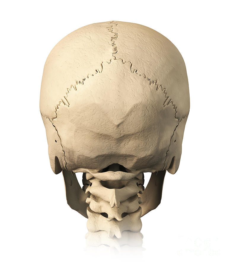
12 photos of the bone of back of skull. The cranium and mandible was exported from ct data. The human skull serves the vital function of protecting the brain from the outside world, as well as supplying a rigid base for muscles and soft tissue structures to attach william is a final year medical student in australia who has taught anatomy to tertiary science and medical students since 2010. Such orbitomeatal plane was accepted as an international standard at an anthropological. Learn this topic now at kenhub! * sutures and fontanelles covered in separate video [link: The most important anatomic structures below the anterior cranial fossa are the orbits and the paranasal sinuses. One of the lines extends from the base of the nose near the eyebrows (called the nasion) to a point at the bottom of the back of the head (called the inion). This article describes the anatomy of the skull, including its structure, features, foramina and contents. The human skull is divided into two major sections ⦁ the temporal process is directed back and laterally to connect to the zygomatic process of the temporal bone. This is a model of the human (homo sapiens) skull. It offers protection to the brain, eye balls, inner ears, and nasal passages. The brain and the front office.each has its own challenges and features.brain skull (lat.
In order to be light, the skull is made up by flat and irregular bones, and has hollow spaces called the sinuses back of skull anatomy. The human skull serves the vital function of protecting the brain from the outside world, as well as supplying a rigid base for muscles and soft tissue structures to attach william is a final year medical student in australia who has taught anatomy to tertiary science and medical students since 2010.


0 Komentar